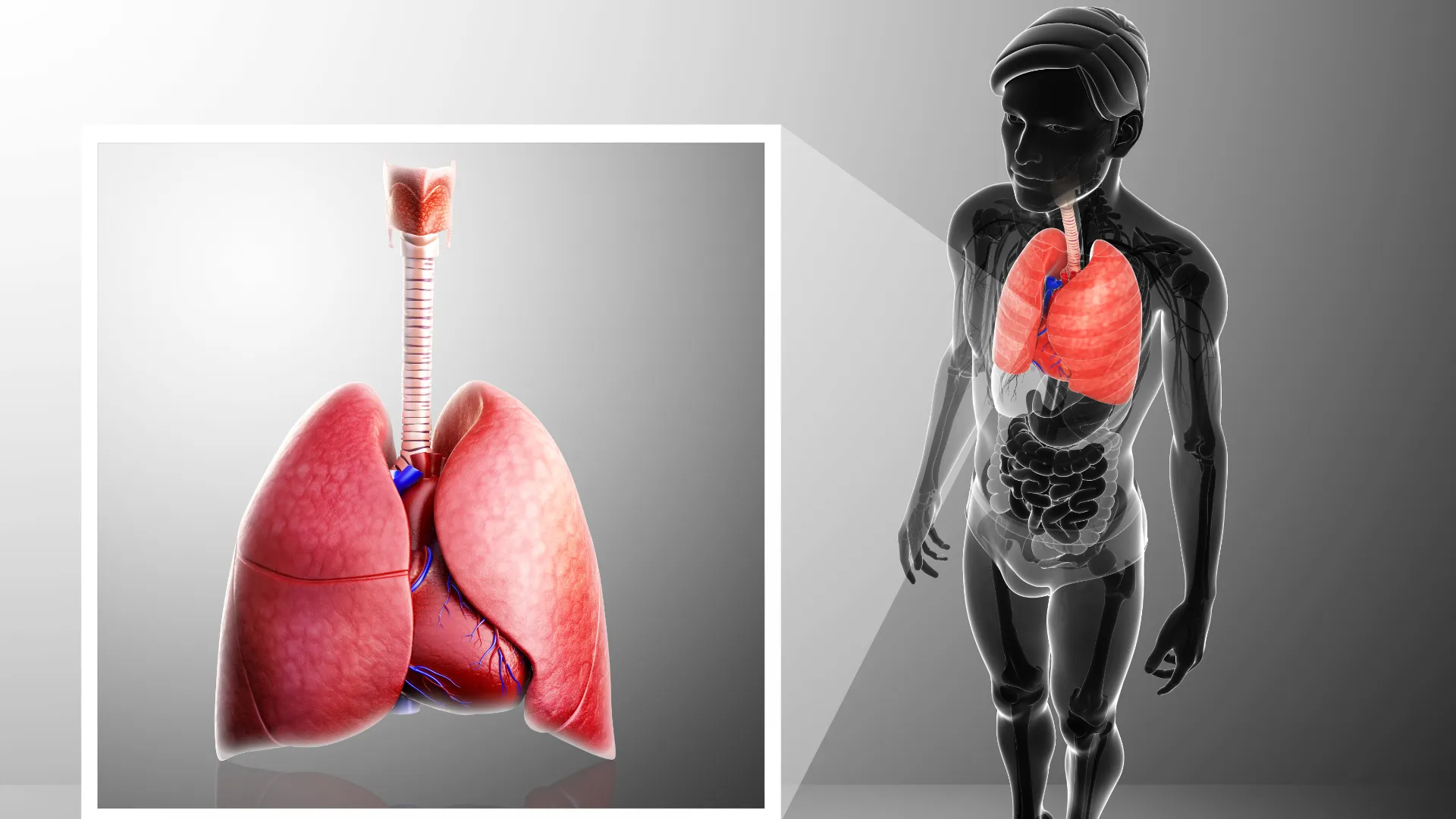
Immunohistochemistry (IHC) is a valuable tool in the diagnosis of epithelioid mesothelioma, a type of cancer that affects the mesothelial cells lining various organs, most commonly the pleura (the lining of the lungs). IHC helps pathologists differentiate epithelioid mesothelioma from other cancers and non-cancerous conditions, as it allows for the detection of specific protein markers associated with mesothelioma. Here are some key steps and markers used in immunohistochemistry for diagnosing epithelioid mesothelioma:
- Tissue Sample Collection: The process begins with the collection of a tissue sample, typically obtained through a biopsy or surgical resection. A tissue sample with suspected mesothelioma cells is essential for diagnosis.
- Fixation and Embedding: The tissue sample is fixed in formalin and embedded in paraffin to preserve the tissue structure for microscopic examination.
- Sectioning: Thin sections (usually 3-5 micrometers thick) of the tissue sample are cut and placed on glass slides. These sections will be used for the immunohistochemical staining.
- Immunostaining: Immunohistochemistry involves the use of specific antibodies that can bind to particular proteins or antigens present in the tissue sample. In the case of epithelioid mesothelioma, several markers are commonly used:a. Calretinin: Calretinin is a calcium-binding protein that is highly specific for mesothelial cells. Strong calretinin staining is often seen in epithelioid mesothelioma.
b. Cytokeratin: Cytokeratins are intermediate filament proteins found in epithelial cells. Epithelioid mesothelioma cells typically express cytokeratin markers, such as AE1/AE3 or CAM5.2.
c. WT-1 (Wilms Tumor 1): WT-1 is a transcription factor expressed in mesothelioma cells. Positive staining for WT-1 is a common finding in epithelioid mesothelioma.
d. D2-40 (Podoplanin): D2-40 is a marker for lymphatic endothelial cells. It can help distinguish mesothelioma from lung adenocarcinoma, which may have similar morphological features.
e. Ber-EP4: Ber-EP4 is another marker that can help differentiate mesothelioma from adenocarcinomas of other origins, such as lung or breast.
- Microscopic Examination: After immunostaining, a pathologist examines the stained tissue sections under a microscope. The presence or absence of staining for these markers, along with the morphology of the cells, is used to make a diagnosis.
- Differential Diagnosis: Epithelioid mesothelioma can be challenging to distinguish from other cancers, especially adenocarcinomas. IHC helps pathologists differentiate mesothelioma from other malignancies based on the pattern of staining and morphology.
It’s important to note that while immunohistochemistry is a valuable tool in diagnosing epithelioid mesothelioma, it is often used in conjunction with other diagnostic methods, such as imaging studies and clinical history, to make a comprehensive diagnosis. Additionally, IHC results should be interpreted by experienced pathologists who are familiar with the nuances of mesothelioma diagnosis.
